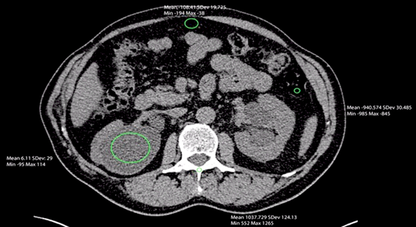
One of the most encountered CT artifacts, beam-hardening artifact, affects the measurement of radiodensity. Polychromatic energies comprise the conventional CT X-ray. ĬT artifacts can affect Hounsfield unit measurements. These factors need standardization to help make the HU a reliable diagnostic measurement tool. The type of reconstructing algorithm, design of the CT, and X-ray kilovoltage, were the most important factors identified. Early studies showed the HU to be dependent on the various CT parameters. Different X-ray beam energies will result in different tissue absorption and hence, different HUs. This linear transformation of the original linear attenuation makes the Hounsfield scale a relative scale, rather than absolute. The Hounsfield unit was named after Sir Godfrey Hounsfield, recipient of the Nobel Prize in Physiology or Medicine in 1979, for his part in the invention of CT, as it had immediate recognition as a revolutionary diagnostic instrument. More dense tissue, with greater X-ray beam absorption, has positive values and appears bright less dense tissue, with less X-ray beam absorption, has negative values and appears dark. The linear transformation produces a Hounsfield scale that displays as gray tones. The upper limits can reach up to 1000 for bones, 2000 for dense bones like the cochlea, and more than 3000 for metals like steel or silver. The Hounsfield unit, also referred to as the CT unit, is then calculated based on a linear transformation of the baseline linear attenuation coefficient of the X-ray beam, where distilled water (at standard temperature and pressure) is arbitrarily defined to be zero Hounsfield Units and air defined as -1000 HU. The physical density of tissue is proportional to the absorption/attenuation of the X-ray beam. The absorption/attenuation coefficient of radiation within a tissue is used during CT reconstruction to produce a grayscale image.

The Hounsfield unit (HU) is a relative quantitative measurement of radio density used by radiologists in the interpretation of computed tomography (CT) images.


 0 kommentar(er)
